The Impact of Ligament Tears on Joint Contact Mechanics in Progressive Collapsing Foot Deformity: A Finite Element Study
Patients with longstanding progressive collapsing foot deformity often develop osteoarthritis of the ankle, midfoot, or hindfoot joints, which can be symptomatic or lead to fixed deformities that complicate treatment. The development of deformity is likely caused by ligament degeneration and tears. However, the effect of individual ligament tears on changes in joint contact mechanics has not been investigated.
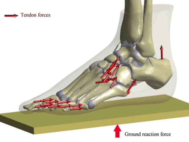
A validated finite element model of the foot was used to compare joint contact areas, forces, and pressures between the intact and collapsed foot, and to evaluate the effect of individual ligament tears on joint
contact mechanics.

Ultrasound Shear Wave Elastography of the Anterior Talofibular and Calcaneofibular Ligaments in Healthy Subjects
Pictured above: CFL was stressed using the talar tilt test, with the orthopedic surgeon applying a medially directed force to invert the heel, while the other hand stabilized the tibia. Note the DataLog Boot Screen myometer used to ensure that 100 N of external force was applied to each stressed ligament.
Most sprained lateral ankle ligaments heal uneventfully, but in some cases the ligament’s elastic function is not restored, leading to chronic ankle instability. Ultrasound shear wave elastography can be used to quantify the elasticity of musculoskeletal soft tissues and may serve as a test of ankle ligament function during healing.

Mechanical Effects of Lag Screw Retightening in a Simulated Hindfoot Arthrodesis Model
Pictured above: Experimental setup. (A) Load cell, (B) 8.0-mm partially threaded cannulated screw, (C) load cell and washers interposed between talus and calcaneus bone cylinders. The screw was advanced from right (calcaneus) to left (talus) over a guidewire (far left) to simulate subtalar arthrodesis.
Nonunion following hindfoot arthrodesis may be caused by failure to maintain compression at the arthrodesis site. The ability of lag screws, commonly used in arthrodesis, to maintain compression in hindfoot bones has not been well characterized. The aim of this work was to quantify the stress relaxation response of hindfoot bone with initial and repeated compression with a lag screw.