
Osseosurface Electronics: The First Permanent Implanted Sensors for Bone Health Data
Advances in bioelectronics have provided important insights into parts of the body that had been largely unexplored. However, the musculoskeletal system has remained an understudied area, in part because of the difficulty of stabilizing sensors on bone and obtaining long-term, reliable signals through a large, soft-tissue envelope.
In 2021, the Biomaterial Lab introduced the first-ever permanent, implantable bone sensors: "osseosurface electronics," created in collaboration with UArizona engineers and other scientists.
These tiny, wireless and battery-free devices include sensors for multiple measures of bone health data as well as LEDs with therapeutic and research applications. The devices permanently bond to bone using a novel calcium ceramic coating.
Advancing Polymer Scaffolding for Bone Repair and Cartilage Generation
The material of choice for bone repair is natural bone – either a patient's own bone tissue or that of someone else (cadaver bone). A patient’s own bone supply is limited, and cadaver bone is less ideal due to the processing needed to minimize the risk of disease transmission.
Metal implants, while readily available, can lead to bone loss and bone density reduction when left in place.
Our researchers are experimenting with physical and chemical properties of a variety of materials and compounds, optimizing performance, overcoming limitations and advancing promising developments in biomaterials musculoskeletal repair.
Repairing Long Bone Segment Defects Using Stem Cells and Polymer Scaffolds
Today there are no clinical treatments that provide an ideal, long-term solution for repairing long bone segment defects. Many patients require additional surgery to induce healing. Patients that heal from these injuries do not always return to pre-injury activities. Failure to heal after multiple surgeries may necessitate amputation.
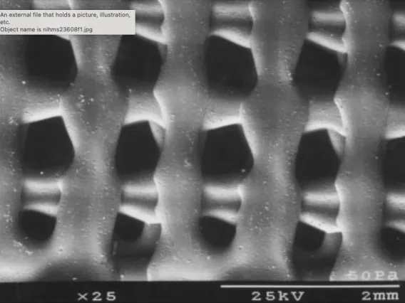
Bone graft material from a patient is limited and may result in morbidity at the donor site. As an alternative solution, the Biomaterials Lab has used 3D-printed scaffolds that mimic biology to facilitate rapid bone bridging in critical-sized defects.
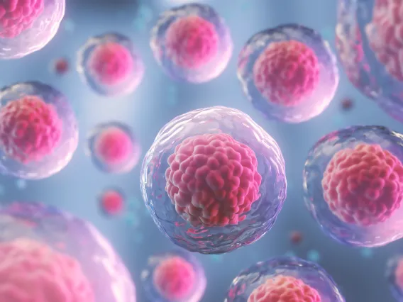
The 3D printed polymer scaffolds we have developed can be combined with adipose derived stem cells, which has been shown to promote rapid bone formation across large defects.
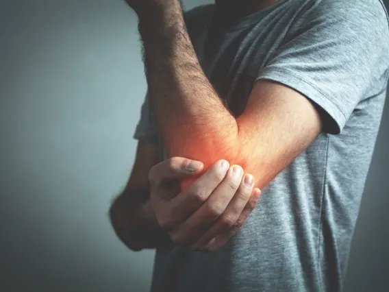
Developing a New Way to Repair Elbow Fractures for Stronger Plate Fixation
A novel method for treating elbow fractures developed at the University of Arizona Department of Orthopaedic Surgery has become part of clinical practice since it was presented at the Orthopedic Research Society annual meeting in 2010 and published in the journal Orthopedic Trauma in 2012.
The technique was developed for fractures of the olecranon – the scoop-shaped part of the elbow joint at the top of the ulna, one of the forearm bones.
By augmenting plate attachments with sutures that transfer a portion of biomechanical forces to the triceps, researchers were able to significantly increase the strength of the fixation. This minimizes the chances of complications when treating fractures with small bone fragments and in patients with osteoporosis.